Rumen Health Technical Guide
Leading ruminant experts have written the Rumen Health Technical Guide. This resource is free to veterinarians, nutritionists and producers, request a copy today.
 Get a copy
Lower Gut Biology
Knowledge of ruminant lower gut biology is essential for understanding the functioning of the ruminant digestive system as a whole and its influence on the ruminant physiology.
What are the different sections of the ruminant lower gut and the functions associated with the ruminant digestive system?
The ruminant lower gut comprises several sections, whose biological specificities (cell diversity, mucus composition) account for the functional differences of each portion of the ruminant lower gut and their role in the ruminant digestive system.
- Small intestine: made up of three sections (duodenum, jejunum and ileum), it measures 20 times the length of the animal. Thanks to secretions from the pancreas and gallbladder, the small intestine handles most of the digestion process in ruminants, as well as absorbing nutrients from the ruminant lower gut lumen into the blood and lymphatic systems (University of Minnesota, 2021).
- Large intestine: this consists of the caecum and the colon. The caecum breaks down certain still undigested fibers, while the colon, the last part of the digestive tract before the rectum, carries undigested food but above all allows water to be absorbed (University of Minnesota, 2021).
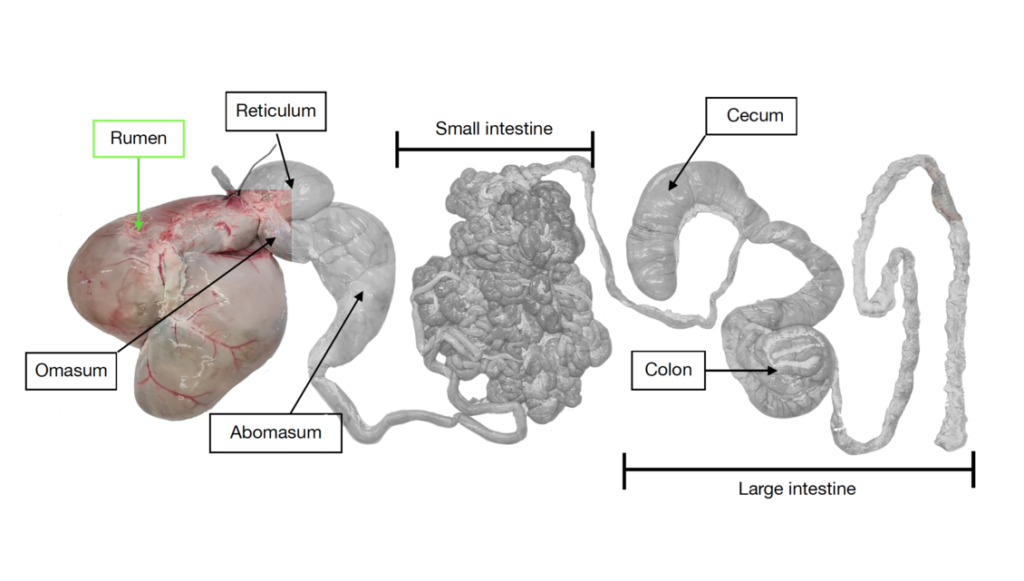
The different parts of the ruminant digestive system (Pain S., 2021)
What are the characteristics associated with ruminant lower gut biology?
The epithelium (or cell wall) of the ruminant lower gut is very different from that of the rumen. It is a specific characteristic of ruminant lower gut biology. This epithelium enables the lower gut to fulfil its double function of absorbing nutrients and acting as a protective barrier (pathogenic and toxic microorganisms), which are key roles in the ruminant digestive system.
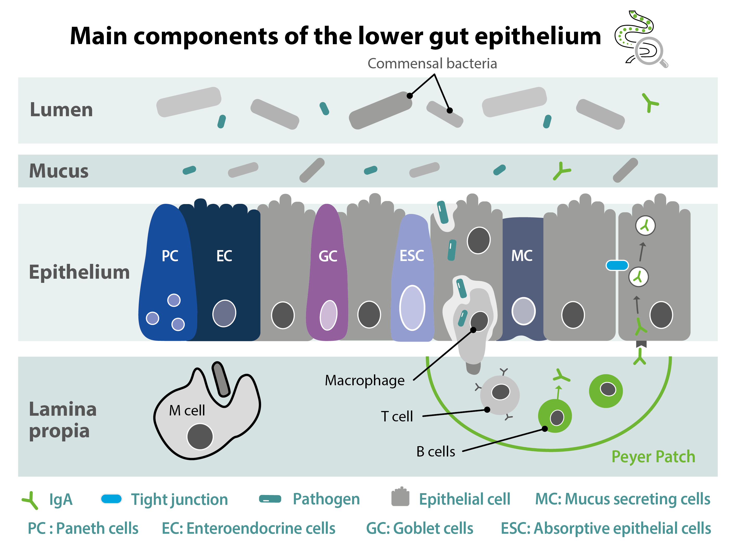
While the rumen has a stratified squamous epithelium with 4 layers, the ruminant lower gut has a single layer of specialized cells (Garcia M. et al., 2017), including:
- Absorptive epithelial cells (ESC): their function is to ensure the absorption of nutrients; the exchange surface is optimized by the presence of ciliated structures on the surface of the cells (Garcia M. et al., 2017).
- Mucus secreting cells (Goblet cells, MC): secrete a protective mucus that lines the intestinal wall (Garcia M. et al., 2017).
- Enteroendocrine cells (Paneth cells, PC): located in the intestinal crypts, they help regulate resident microbial populations by secreting antimicrobial peptides into the small intestine (Steele M.A. et al., 2016).
- Immune cells (M cells): these facilitate the transport of antigens and microorganisms from the intestinal lumen to the underlying intestinal cells (dendritic cells, lymphocytes) (Garcia M. et al., 2017).
These cells are closely interconnected by the mucus layer that covers their apical surface, as well as by specific junctions (tight junctions – TJs, adherens junctions – AJs, and desmosomes) which are essential for maintaining the physical structure (Kocot A.M. et al., 2022).
The diversity and complementarity of these different cell types making up the wall of the ruminant lower gut enables to perform many of the physiological functions required by the ruminant digestive system.
However, it is also essential to consider the microbial diversity and density of the ruminant lower gut to appreciate the full ruminant physiology of this important part of the ruminant digestive system. If you would like to find out more about the ruminant lower gut microbiotaGlossaryView allMicrobiota
A microbiota is the whole of the ecosystem (bacteria, yeast, fungi and viruses) living in a specific environment. Intestinal microbiota was previously called “intestinal flora.”, click here.
